All posts by analyticalansw

I’m Looking Through You : Q&A

In this webinar, we look at the capabilities of TEM and its methodologies. The Q & A from the webinar provides a brief FAQ about this type of analysis.

Q: How large a field of view can you prepare for TEM?

A: While we cannot image the entire prepared sample at once, we typically prepare TEM sections by FIB with an electron transparent region that is about 10 µm in length. We actually prefer an area that is smaller than that just to maintain mechanical stability, but that’s the typical limit of FIB preparation. In a TEM sample that is prepared by ultramicrotomy, an ultrathin sample can be up to 2 or 3 mm in diameter.


Q: Can any material be prepared for TEM?

A: Most likely not. There are some restrictions around what we can put into the transmission electron microscope. Again, the most important restrictions are that it needs to be vacuum compatible and that it needs to be able to stand up to the FIB preparation if we go the FIB route. However, some polymer samples that may be vacuum compatible might be ‘sectionable’ with our ultramicrotome but may degrade under an electron beam. Some could even be too soft to section by ultramicrotomy. There are many, many materials that can be used for TEM, however.


Q: Can TEM be used to measure crystallinity index as XRD?

A: In a single crystal sample, we get well-resolved diffraction patterns that we can use to measure lattice parameters and similar aspects of crystallinity.


Q: Is EDS analysis in the TEM or STEM more time consuming than in the SEM?

A: The answer to that question is no. It typically takes about the same amount of time once you have your sample in the instrument to actually generate the EDS analysis.


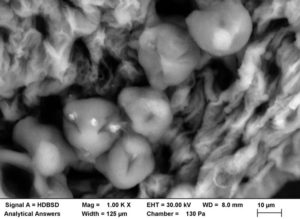
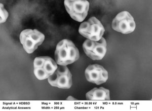
Q: Can you do grain boundary imaging and analysis in the TEM?

A: Typically, the answer to that question is yes. Again, it can be somewhat sample dependent, but the huge advantage in the TEM or STEM is that the sample is so thin that there is virtually no spot blooming compared to bulk EDS analysis in the SEM, allowing us to perform EDS analysis on a nanometer rather than micron scale. So if you have a problem along those lines, again, we would certainly encourage you to get in touch with us and we can see about designing an analysis to address that.


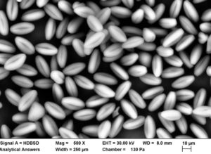
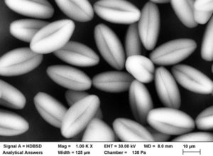
Q: Can you do grain or particle size analysis from TEM images?

A: Again, the answer to that, in general, is yes. However, you will need to contact us to confirm.


Q: Can you make a TEM sample from a four-inch wafer?

A: Yes. On a four-inch wafer, if there are small structures or small defects that we need to find, the capability exists to drive to those positions and then excise those regions of interest from the wafer as lift-out samples and prepare them for subsequent TEM analysis.


Q: What is the difference between high resolution TEM and STEM?

A: STEM in its name implies that you are scanning the beam: Scanning Transmission Electron Microscopy. In that technique, we would form a finely focused probe beam, step it across the sample in a raster fashion to generate the image (as is done in an SEM) but we take advantage of the higher beam voltages and different electron optics to provide additional signals and information about samples, as compared to SEM. In a high resolution TEM image or TEM in general, the beam is not scanned; we rely on the electron optics to form a global image of the sample, much as you would in a light microscope.

Small Spot ESCA : Q&A

The above webinar discusses analyzing surface chemistry on micro-devices and its methodologies. Here you can examine a Q & A from the webinar of FAQs about this type of analysis.

Q: Does surface roughness have an effect on depth profiles?

A: Surface roughness does indeed have an effect on depth profiling. But with our new instrumentation, our ability to rotate a sample at multiple points coupled with our better charge neutralization helps to mitigate the effects that you would see from surface roughness. It may cause your sample to sputter etch at a bit slower rate than expected, but rotating will help decrease the effects that we see from that. Charge neutralization also helps to mitigate the effects one sees. So yes it’s possible, but there are caveats.


Q: How deep can you profile? How long does it take to sputter?

A: That depends on the sputter rate of the material under analysis. Again, we do a quality check four times a year on our instrumentation. And we know that silicon dioxide sputters at a rate of 10 nanometers per minute in our instrumentation. We have parameters that can increase or decrease that sputter rate and make it much faster or much slower, but if that’s a benchmark, it’s limited. Thus, with the 10-nanometer sputter rate, it just limits how deep we can get into the sample.

And some materials may sputter faster or slower, so it all depends on how much time you want to spend to get, say, a micron into the surface.

Something to keep in mind as you’re sputtering past a micron is that interfaces may be intermingled as you’re getting deeper into the surface because of the crater that’s being created. It depends on the data that can be collected, as well as the electronics, and the set-up of the instrumentation. Those are all factors to keep in mind as you’re sputtering one to two to three microns into a sample. Time is a big factor– how long do you want to spend to sputter a micron or more into your sample.

It’s possible, and we can do it. But once you get into the two to three micron range, there are several things to keep in mind. You may see artifacts as we sputter that deep into the sample. As long as you remember that there may be artifacts and that that data may have to be taken with a grain of salt, you can sputter as deep as you want, as long as you have the time to do it.


Q: Can you give me an example of when weight percentage analysis would be needed?

A: Weight percentage analysis is often needed when a client comes in with an idea of a product that’s been on the surface. It’s not always possible, but sometimes they say, hey, we know the weight percent of this product. Can we see some of that as we analyze our sample? So let’s say we have a particular product of poly dimethyl silicone, and can we see that reflected on our sample. Sometimes that can be calculated to weight percent that can be found. Weight percentage can also be useful or beneficial if you’ve tried to create a product similar to a competitor’s and you want to see if that works out. Sometimes that can be back calculated. Sometimes that comes with a caveat.


Q: How can you produce maps and line scans similar to what you showed in your depth profiling system?

A: A map is actually made up of multiple points so we analyze each point and software will combine the data to give it to you. So you are losing some sensitivity to data because it is a multidimensional analysis. We recommend doing a point analysis to get your overall composition. We take multiple points, and just like a pixelated picture the software combines all those pixels.

Fast Mapping : Q&A

The above webinar discusses the capabilities of fast mapping and its applications. Here you can read the Q & A from the webinar for a series of FAQs on the topic of fast mapping.

Q: Do you have to define elemental windows before starting the map?

A: The answer is no. Because you’re getting an entire spectrum from each pixel in the map, you don’t have to define anything ahead of time or after the fact – that’s locked in. You can always go back and change the elements you’ve mapped for, so that way you can dynamically change it as needed. You can even add elements partway through. You can subtract them if you find that they don’t make sense because they shouldn’t be there. But again, if you don’t agree with the spectrometer, you can always check your work by extracting synthesized spectra containing the same elements, and verifying that any decisions that you came to are actually accurate and correct.


Q: Is this method able to be used for polymer or non-metallic materials?

A: The answer is yes. You can certainly use this to look at polymeric materials. As with all SEM work on polymers, you typically have to coat the sample with a conductive coating. Otherwise, you will end up with charging problems because the sample cannot dissipate the charge from the electron beam.


Q: How deep into the sample does the beam penetrate?

A: The beam penetrates anywhere from one up to tens of microns, depending on the density of the material and the energy of your beam. The scale in the webinar example is 10 microns in the lateral dimension, but the same holds true in the vertical direction. Thus, when you’re hitting a low atomic number material like aluminum with a 30 kilovolt beam, most of the information will come from the outermost few microns but you can still get some data from much deeper into the sample surface. So if you’re looking for very sensitive detection, you can still pick up those very weak traces of elements when they’re coming in from a distance. As you can see with the iron comparison, the penetration depth is much smaller because of the higher atomic number of that material. And so you also get a smaller analytical volume with most of the information coming from the top 2 microns. But you can still get some data from as deep as 5 microns, but it’s not the highest intensity data that’s coming from the fringes of those “spot blooms” where the e-beam spreads out into the material.

Beaches: East vs. West – a study in sand


Now that summer has drawn to a close, we’re taking a look back at some beaches that we visited this summer. Noting that the sand looked and felt different from beach to beach, we sampled a bit of sand from each – and later, from beaches on the west coast as well. The east coast samples were collected at various times through the summer. The California samples were collected in August and September on a drive up the coast from Long Beach to Tiburon.

Just as the character and feel of each of these beaches is different, the sand samples also look quite different. But how different are they really? Take a closer look with a special optical microscope that allows us to acquire a series of images in multiple focal planes and combine them into a single image with a very large focal depth.

Each of the Extended Focus images are a composite of between 90 and 125 optical microscope images taken under comparable lighting conditions. Extended Focus mode allows us to keep all features in these 3-dimensional sample in focus through a depth of more than 1 mm. All of the sands were imaged at an original magnification of 35X so that grain sizes and material “mixtures” could be directly compared.

Turkey Feathers: Intricate Structures

Here, we’re featuring the structures of turkey feathers. More specifically, some optical and electron microscope images of the intricate structure of a portion of a tail feather donated by a wild turkey (Meleagris gallopavo) from a large flock living on the Danvers-Wenham, MA town line.


Wild Turkey tail feather 10x
Wild Turkey tail feather 20x
Tail Feather Zstack, Extended Focus Mode, 60x

The large central shaft (rachis) is visible near the top of the lowest magnification optical (above) and electron microscopic (below) images, with the array of barbs branching off it. The barbs form the two vanes of the feather, one on each side of the rachis. Branching off the barbs are the barbules. The tiny hooks that hold the barbules together are called barbicels, and are clearly visible in the SEM images below.

61x
100x
500x

At low magnification (original magnifications are shown), optical images were taken using a stereo zoom microscope. At higher magnification however, the 3-dimensional nature of the feather structures make it difficult to capture a fully “in-focus” image. One way to solve that problem is to acquire a composite (in this case, approximately 100) images at different focal planes (Extended Focus Mode). The resulting image is in focus throughout the entire depth of the features present in the sample. The same portion of the feather was then mounted for viewing in the Scanning Electron Microscope (SEM), and a thin (approximately 2 nm) of gold-palladium metal was sputtered onto the sample to provide a conductive sample surface to minimize charge buildup from the incident electron  beam. Because the wavelength of electrons is considerably shorter than that of visible light, the electron microscope provides significantly greater depth of field and depth of focus than the optical microscope.

Analytical Answers, Inc. to Bring Materials Analysis Expertise to Advanced Manufacturing Convention

FOR IMMEDIATE RELEASE

Analytical Answers, Inc., a Woburn, Massachusetts-based technical consulting firm and service provider which helps advanced manufacturing firms monitor their processes and solve R&D and production problems, will be exhibiting at this year’s Advanced Manufacturing convention June 12-14 at the Javits Convention Center in New York City.

We are excited to be discussing solutions to some of advanced manufacturing’s toughest quality and analysis challenges, using technologies available in their laboratory that are normally only found in large corporations or research facilities, combined with the unique expertise of their hands-on team of scientists.

Five-Minute Material Analysis Update Available

The scientists and staff at Analytical Answers welcome existing clients to their booth and invite new clients to stop by and discover the many ways that the company can help explore micro-to-nano level surface and bulk composition and structure, wear, and failure mode analysis. This year they have also made significant additions to their extensive repertoire of optical, x-ray, electron, and ion-based analytical capabilities. They are now offering Broad Ion Beam Milling (BIB) and small spot ESCA, also known as X-Ray Photoelectron Spectroscopy (XPS). They note that “BIB rapidly sections through large (3mm+) areas of materials, including some that often can’t be cut by FIB, ultramicrotomy or polishing techniques. Small Spot XPS provides a surface analysis with a lateral resolution of 30µm, compared to a spot size greater than 400µm previously available with “standard” ESCA.” Adding these techniques for sectioning and analysis expands the range of materials that can be analyzed and enables surface analyiss (1-4 atom layers, with ion sputtering provide depth proviling capabilities) with significantly better spatial resolution.

The company has added these capabilities to extend their ability to analyze customer materials with more advanced nano-level analysis including:

  • Comparing oxidation depth across samples
  • Checking coating and lubrication thickness and composition
  • Examining subsurface material composition by ion sputtering and scanning
  • Checking chemical consistency of materials, for example differentiating sulfide from sulfite
  • Preparing improved samples to visually or chemically analyze microscopic structures, fillers, or inclusions (such as in/on circuit boards or in polymers such as catheters or extruded materials).

The new capabilities at Analytical Answers, Inc. extend the abilities of their scientific staff, who have a combined 100-plus years of experience. They will now be able to work with clients to examine surface features as small as 30µm on conductive and insulating materials, yielding greater information on isolated anomalies, and material characteristics that can only be revealed with this more finely-focused probe size.

Specific Benefits

Manufacturers producing medical equipment such as surgical mirrors, with coatings consisting of multiple nanometer-thick layers will benefit from the detailed analysis of their product using XPS, and the ability to check production samples against FDA requirements. Clients can also verify the quality of new materials such as graphene using XPS. BIB allows rapid sectioning of previously problematic materials such as soft polymer materials often found in medical devices, or other composites and coatings that can be distorted by conventional preparation techniques, facilitating analysis by other techniques such as Scanning Auger Microanalysis or Scanning Electron Microscopy.

A Corporate Lab for a Diverse Range of Manufacturers

Only the largest corporate laboratories have the capabilities Analytical Answers has to offer its clients. Analytical Answers is pleased to also work with these companies, when priority scheduling is required but unavailable at their in-house laboratories, or when additional analytical capacity is needed. They are also at the Advanced Manufacturing exhibit to welcome smaller manufacturing companies who don’t routinely use analytical laboratory services but have occasional manufacturing problems or quality issues that might benefit from analysis. The benefits could be improved product or process quality better understanding how  processes are actually working and what materials or characteristics could be further improved.

Preparing for Future Production

In the fast-paced technology development environment of advanced manufacturing, future-oriented companies will seek the data about their products and their composition that Analytical Answers routinely provides. Quality assurance testing can be moved from operational pass-fail to production sample analysis which verifies that product margins are being maintained. Analytical Answers provides information which can help advanced manufacturing companies start planning improvements to their product and production techniques ahead of the curve.

A Summary of their New Capabilities

Broad Ion Beam Milling and Small Spot ESCA (Electron Spectroscopy for Chemical Analysis, or X-ray Photoelectron Spectroscopy – XPS).

  1. Broad Ion Beam Milling (BIB) rapidly sections through large (3mm+) areas of materials, including some which couldn’t be cut by other methods such as brittle materials, soft polymers, or materials with inclusions or fillers of varying hardnesses.
  2. Small Spot ESCA provides a surface analysis spot size of 30µm, along with surface sputtering and depth profile capabilities. The improved analysis area provides an order of magnitude improvement in spatial resolution, compared to previously available ESCA instruments.

These new capabilities are in addition to a large menu of existing analysis options, available here.

Come visit Analytical Answers at booth 559 to find out more! Of course, you can also call, email, or contact them through their Web site, www.analyticalanswersinc.com.

***

Analytical Answers is a consulting and technical services company providing microanalysis, surface characterization, and failure analysis. With over 100 years of combined experience among our scientists, and instrumentation capabilities usually only found in corporate or research laboratories, we provide resources and expertise to translate data into solutions.

Pollen

The nice spring weather has come and gone. With that nice spring weather also came pollen…everywhere! Although it is often seen as a nuisance for triggering allergies, pollen is very interesting to look at magnified. You may be surprised at how different various pollen types look.


The familiar yellow haze on our cars and everywhere else can be annoying. But the variation in the appearance of different types of pollen at higher magnifications can be surprising.

Pollen close up:


Empress Tree Flower Pollen – This tree (Paulownia tomentosa) produces a small grain size pollen with a “bean” or “seed” shape and longitudinal folds.

Empress Tree Flower Pollen 2 – While the wall surfaces appear smooth at lower magnifications, a mottled appearance begins to appear at higher magnification.
Gala Apple Blossom Pollen – The pollen grains shown here are still seated on the anther surface. They are quite large with a smooth wall surface, and appear almost “fluffy” with only a few folds.
Pink Azalea Pollen – These are relatively large pollen grains with a faceted wall structure.
Pink Azalea Pollen 2 – At higher magnification, the wall surface has the appearance of connected groups of dimpled facets with a “stitch” that connects the facets.
Russian White Lilac Pollen – These medium-size grains have a mottled wall surface with one or two folds.

Russian White Lilac Pollen 2 – At higher magnification the folds on some grains actually look like holes. The wall surface texture is also clearly visible.


Ticks: The More You Know

The image of the month is a false-colored secondary electron (SE) image of the mouth parts of a tick, taken using a Scanning Electron Microscope (SEM). The subject is most likely a male deer tick (shown below in the black-and-white SE image), commonly found in Massachusetts and across the northeast – and a prime carrier of the bacteria that causes Lyme disease, Borrelia burgdorferi (B. burgdorferi).

In February of 2016, the Centers for Disease Control announced that a new species of this bacteria, B. mayonii also causes Lyme in people. And now a new tick-borne virus that is on the rise – Pawassan – was a recent topic of discussion on National Public Radio’s On Point program. Pawassan can cause paralysis and death in humans, representing a new threat in what has been described as the worst year for ticks in recent history – largely because of the wet conditions nationwide over the winter and spring.

The false-coloring was collected by acquiring multiple images using a solid state backscatter detector that allows the user to select specific parts of the detector for image acquisition. Using an image processing program, colors can be assigned to specific images that are then combined to produce a natural-looking color scheme. Both the false-color and ‘normal’ images were collected using special lower-vacuum conditions to avoid drying and other artifacts that occur under the normal high-vacuum environment of the SEM.

The Science of Sundaes


It’s a hot, humid summer day; the sun is blazing down making waves of heat bounce off the pavement, the air is thick with moisture and there’s no sign of a breeze anytime soon. There are many ways to cool off in times like these: having a cold glass of lemonade, taking a refreshing dip in a pool, or even just staying inside planted next to the AC. But the best way to cool off is with a frozen dairy treat. In other words, ice cream.

I’m sure most people can recall countless hot days like this, and the giant wave of relief when the sound of the ice cream truck appeared in the distance, the anticipation growing as the sound grew closer. Although there are always numerous options of ice cream and popsicles, there is nothing more satisfying than a traditional ice cream sundae. And a crucial part of ice cream sundaes is the whipped cream – and the best whipped cream doesn’t come out of a can, it’s made fresh.

In honor of the summer season (or as I like to call it the “months of sundaes”) we took some images of whipped cream with an electron microscope to reveal the microscopic makeup of this delicious topping. Now that we have a closer look, let’s grab a spoon and dig in to the science behind whipped cream

The magic begins with the whipping process. When liquid cream is being whipped, millions and millions of tiny air bubbles start to form within the liquid. Although this process starts to thicken the cream and make it frothy, this early stage of whipping does not result in the final creamy product that we all know and love. If stopped too early, the bubbles would dissipate and the cream would go back to its initial liquid phase.

However, when the whipping process is continued, the cream begins to thicken more and more to a point where it cannot return to the liquid phase. This occurs because of the fats, called trScanning Electron Microscope image of whipped cream, Boston Analytical Labiglycerides, inside the cream. The triglyceride molecules in milk come with protective membranes, called phospholipids, similar to the shell on an egg. The whipping begins to break down these protective membranes on the triglyceride molecules. When the phospholipids are removed from the triglyceride membrane, they join together and then form protective barriers around the air bubbles. Then, when the air bubbles have their own protective membrane, they stick together and form larger and larger clumps, preventing them from dissipating and stopping the cream from going back to its liquid phase.

This means that whipped cream is technically an emulsion, or air suspended in a liquid and stabilized by fat. This is why whipping cream has to have a higher fat content in order for the whipped cream to form; if it doesn’t have enough fat, the air bubbles will dissipate and the cream will remain a liquid. This process is also similar to the formation of other whipped dairy products, like butter and ice cream.

So now that you know how whipped cream is made and what it looks like on a microscopic level, go ahead and enjoy a sundae topped with this whipped, fatty emulsion and think about all the little air bubbles inside of it as the spoon hits your tongue. And of course, don’t forget the cherry!


Sources:

http://www.thekitchn.com/food-science-how-whipped-cream-81751

https://www.uoguelph.ca/foodscience/book-page/whipped-cream-structure

https://foodretro.com/the-science-of-whipped-cream-and-butter/ 


Drew Killius – A Curious Scientist is the Best Kind

According to Drew Killius, searching for (and finding) answers is the best way to explain how the world works. And when it doesn’t, Killius is probably the guy you want working on the reasons behind a product’s failure.

As Senior Materials Scientist at Analytical Answers, Inc., he’s been asked multiple times about the questions he has to ask on a daily basis. His inquisitiveness once even drove an acquaintance to stridently ask, “Why do you have to know all that stuff?!”

Killius’ answer would likely be – though we didn’t want to ask him the question again – to solve problems, help companies and consumers, and to locate solutions. With a background in chemistry and geology, he chose the sciences as a career. Though he says if he went in a different direction vocationally, it would have been in a skilled field like tradesman, plumber or even airline pilot. Killius still gets to use some of his hands-on fixing skills at AAI when instruments act up or there’s a small machine-shop project to tackle.

Similar to the other scientists at Analytical Answers, Killius has worked on numerous projects that can’t be discussed in detail because of non-disclosure agreements. But when asked about his successes, he points to whitepapers that discuss materials and their composition. These are available at the AAI lab in Woburn and here on the Website: Failure Analysis of a Motorcycle Suspension and The Tale of a Spark Plug.

According to Killius, “Many of the things I work on are high-reliability items that aren’t. This frightens people.”

Ultimately, it’s through the analysis Killius performs on products and their material make-up that helps companies improve devices and eliminate defects. Without the AAI team, there might be more items in use that are prone to failure…and these failures can have significant consequences.

Hypothetically, you only have to think about what might happen if a toothpaste tube’s material was incapable of keeping toothpaste inside under certain environmental conditions. While less serious than some of the cases Killius works on, the toothpaste tube imaginary example could become a messy nuisance. Be assured, his real case studies and projects are much more serious. Yet he has an affinity for the companies and brands he helps.

I use the products that I work on, because I know that the folks who make them give a damn,” said Killius. “It’s all those other guys that I never see that I worry about.”

Outside of the lab, he’s also active and energetic. Aside from his love of photography, Killius enjoys motorcycling, high-end woodworking projects, and he’s even made several telescopes by hand.

Science is a profession that attracts all kinds of people, but the trait the best scientists possess is the same:  A curious nature and the drive to find answers to challenges as yet unsolved.

Drew Killius fits that description to a T.