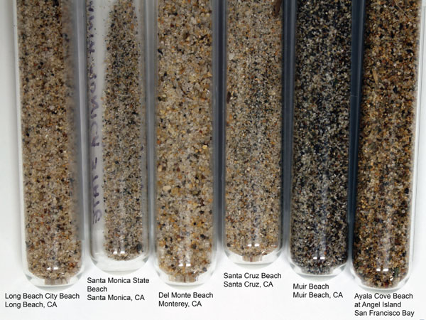
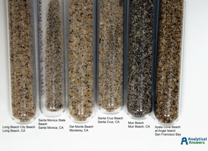
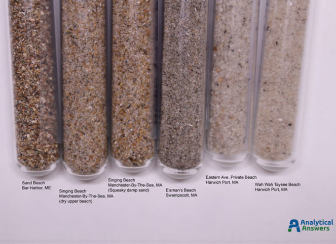
Now that summer has drawn to a close, we’re taking a look back at some beaches that we visited this summer. Noting that the sand looked and felt different from beach to beach, we sampled a bit of sand from each – and later, from beaches on the west coast as well. The east coast samples were collected at various times through the summer. The California samples were collected in August and September on a drive up the coast from Long Beach to Tiburon.
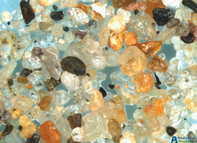
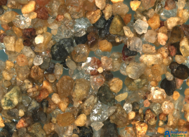
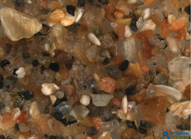
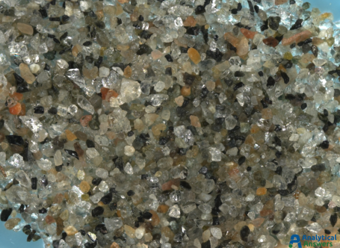
Just as the character and feel of each of these beaches is different, the sand samples also look quite different. But how different are they really? Take a closer look with a special optical microscope that allows us to acquire a series of images in multiple focal planes and combine them into a single image with a very large focal depth.
Each of the Extended Focus images are a composite of between 90 and 125 optical microscope images taken under comparable lighting conditions. Extended Focus mode allows us to keep all features in these 3-dimensional sample in focus through a depth of more than 1 mm. All of the sands were imaged at an original magnification of 35X so that grain sizes and material “mixtures” could be directly compared.
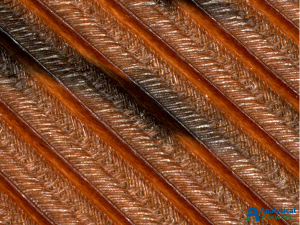
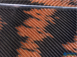
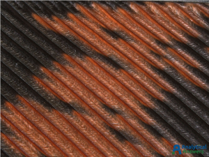
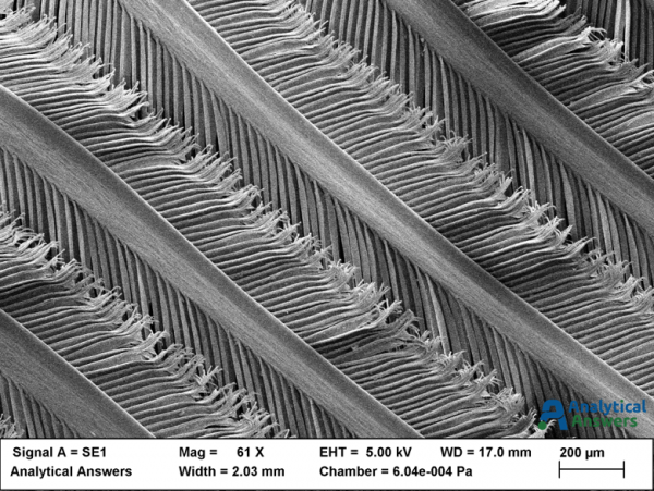
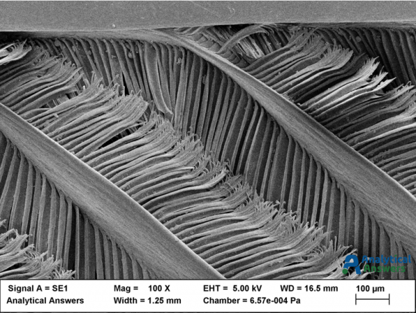
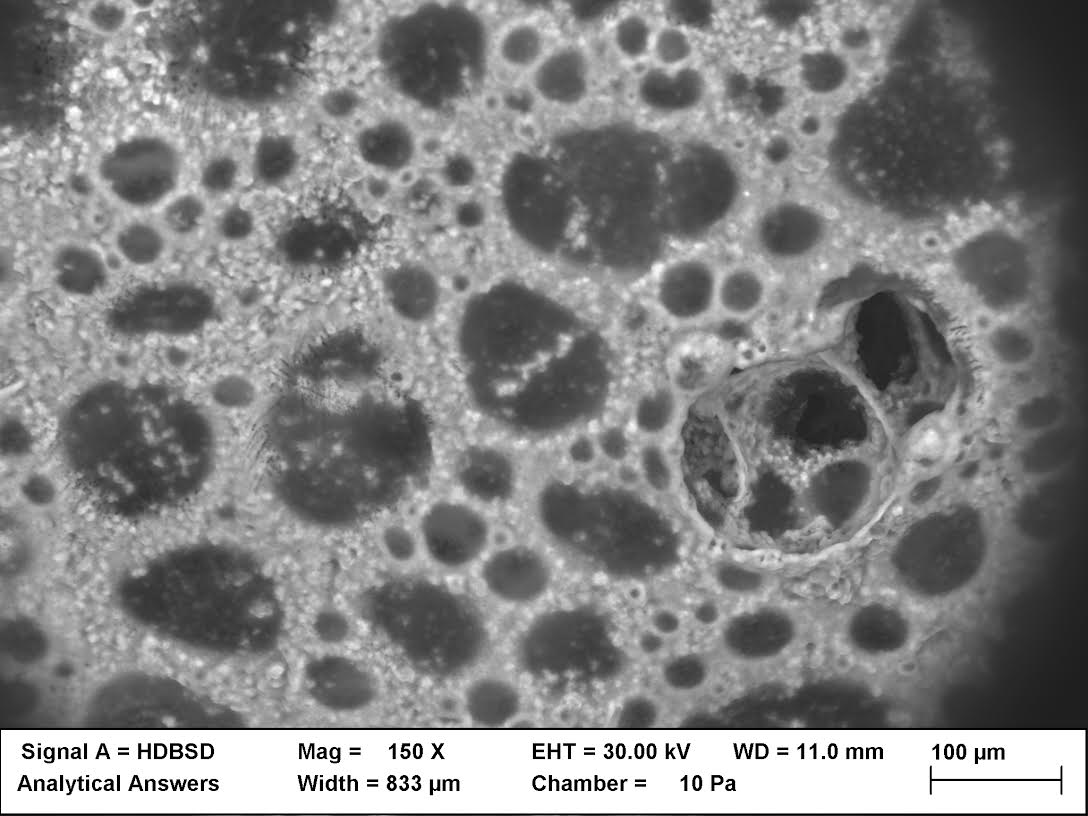
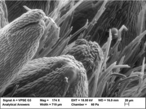
Here, we’re featuring the structures of turkey feathers. More specifically, some optical and electron microscope images of the intricate structure of a portion of a tail feather donated by a wild turkey (Meleagris gallopavo) from a large flock living on the Danvers-Wenham, MA town line.
Wild Turkey tail feather 10x
Wild Turkey tail feather 20x
Tail Feather Zstack, Extended Focus Mode, 60x
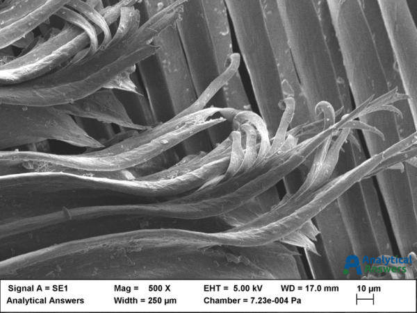
The large central shaft (rachis) is visible near the top of the lowest magnification optical (above) and electron microscopic (below) images, with the array of barbs branching off it. The barbs form the two vanes of the feather, one on each side of the rachis. Branching off the barbs are the barbules. The tiny hooks that hold the barbules together are called barbicels, and are clearly visible in the SEM images below.
61x
100x
500x
At low magnification (original magnifications are shown), optical images were taken using a stereo zoom microscope. At higher magnification however, the 3-dimensional nature of the feather structures make it difficult to capture a fully “in-focus” image. One way to solve that problem is to acquire a composite (in this case, approximately 100) images at different focal planes (Extended Focus Mode). The resulting image is in focus throughout the entire depth of the features present in the sample. The same portion of the feather was then mounted for viewing in the Scanning Electron Microscope (SEM), and a thin (approximately 2 nm) of gold-palladium metal was sputtered onto the sample to provide a conductive sample surface to minimize charge buildup from the incident electron beam. Because the wavelength of electrons is considerably shorter than that of visible light, the electron microscope provides significantly greater depth of field and depth of focus than the optical microscope.
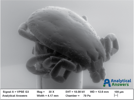
The image of the month is a false-colored secondary electron (SE) image of the mouth parts of a tick, taken using a Scanning Electron Microscope (SEM). The subject is most likely a male deer tick (shown below in the black-and-white SE image), commonly found in Massachusetts and across the northeast – and a prime carrier of the bacteria that causes Lyme disease, Borrelia burgdorferi (B. burgdorferi).
In February of 2016, the Centers for Disease Control announced that a new species of this bacteria, B. mayonii also causes Lyme in people. And now a new tick-borne virus that is on the rise – Pawassan – was a recent topic of discussion on National Public Radio’s On Point program. Pawassan can cause paralysis and death in humans, representing a new threat in what has been described as the worst year for ticks in recent history – largely because of the wet conditions nationwide over the winter and spring.
The false-coloring was collected by acquiring multiple images using a solid state backscatter detector that allows the user to select specific parts of the detector for image acquisition. Using an image processing program, colors can be assigned to specific images that are then combined to produce a natural-looking color scheme. Both the false-color and ‘normal’ images were collected using special lower-vacuum conditions to avoid drying and other artifacts that occur under the normal high-vacuum environment of the SEM.
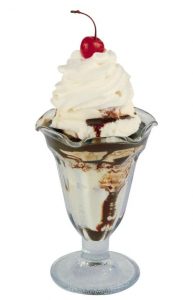
It’s a hot, humid summer day; the sun is blazing down making waves of heat bounce off the pavement, the air is thick with moisture and there’s no sign of a breeze anytime soon. There are many ways to cool off in times like these: having a cold glass of lemonade, taking a refreshing dip in a pool, or even just staying inside planted next to the AC. But the best way to cool off is with a frozen dairy treat. In other words, ice cream.
I’m sure most people can recall countless hot days like this, and the giant wave of relief when the sound of the ice cream truck appeared in the distance, the anticipation growing as the sound grew closer. Although there are always numerous options of ice cream and popsicles, there is nothing more satisfying than a traditional ice cream sundae. And a crucial part of ice cream sundaes is the whipped cream – and the best whipped cream doesn’t come out of a can, it’s made fresh.
In honor of the summer season (or as I like to call it the “months of sundaes”) we took some images of whipped cream with an electron microscope to reveal the microscopic makeup of this delicious topping. Now that we have a closer look, let’s grab a spoon and dig in to the science behind whipped cream
The magic begins with the whipping process. When liquid cream is being whipped, millions and millions of tiny air bubbles start to form within the liquid. Although this process starts to thicken the cream and make it frothy, this early stage of whipping does not result in the final creamy product that we all know and love. If stopped too early, the bubbles would dissipate and the cream would go back to its initial liquid phase.
However, when the whipping process is continued, the cream begins to thicken more and more to a point where it cannot return to the liquid phase. This occurs because of the fats, called triglycerides, inside the cream. The triglyceride molecules in milk come with protective membranes, called phospholipids, similar to the shell on an egg. The whipping begins to break down these protective membranes on the triglyceride molecules. When the phospholipids are removed from the triglyceride membrane, they join together and then form protective barriers around the air bubbles. Then, when the air bubbles have their own protective membrane, they stick together and form larger and larger clumps, preventing them from dissipating and stopping the cream from going back to its liquid phase.
This means that whipped cream is technically an emulsion, or air suspended in a liquid and stabilized by fat. This is why whipping cream has to have a higher fat content in order for the whipped cream to form; if it doesn’t have enough fat, the air bubbles will dissipate and the cream will remain a liquid. This process is also similar to the formation of other whipped dairy products, like butter and ice cream.
So now that you know how whipped cream is made and what it looks like on a microscopic level, go ahead and enjoy a sundae topped with this whipped, fatty emulsion and think about all the little air bubbles inside of it as the spoon hits your tongue. And of course, don’t forget the cherry!
According to Drew Killius, searching for (and finding) answers is the best way to explain how the world works. And when it doesn’t, Killius is probably the guy you want working on the reasons behind a product’s failure.
As Senior Materials Scientist at Analytical Answers, Inc., he’s been asked multiple times about the questions he has to ask on a daily basis. His inquisitiveness once even drove an acquaintance to stridently ask, “Why do you have to know all that stuff?!”
Killius’ answer would likely be – though we didn’t want to ask him the question again – to solve problems, help companies and consumers, and to locate solutions. With a background in chemistry and geology, he chose the sciences as a career. Though he says if he went in a different direction vocationally, it would have been in a skilled field like tradesman, plumber or even airline pilot. Killius still gets to use some of his hands-on fixing skills at AAI when instruments act up or there’s a small machine-shop project to tackle.
Similar to the other scientists at Analytical Answers, Killius has worked on numerous projects that can’t be discussed in detail because of non-disclosure agreements. But when asked about his successes, he points to whitepapers that discuss materials and their composition. These are available at the AAI lab in Woburn and here on the Website:FailureAnalysis of aMotorcycle Suspensionand The Tale of a Spark Plug.
According to Killius, “Many of the things I work on are high-reliability items that aren’t. This frightens people.”
Ultimately, it’s through the analysis Killius performs on products and their material make-up that helps companies improve devices and eliminate defects. Without the AAI team, there might be more items in use that are prone to failure…and these failures can have significant consequences.
Hypothetically, you only have to think about what might happen if a toothpaste tube’s material was incapable of keeping toothpaste inside under certain environmental conditions. While less serious than some of the cases Killius works on, the toothpaste tube imaginary example could become a messy nuisance. Be assured, his real case studies and projects are much more serious. Yet he has an affinity for the companies and brands he helps.
“I use the products that I work on, because I know that the folks who make them give a damn,” said Killius. “It’s all those other guys that I never see that I worry about.”
Outside of the lab, he’s also active and energetic. Aside from his love of photography, Killius enjoys motorcycling, high-end woodworking projects, and he’s even made several telescopes by hand.
Science is a profession that attracts all kinds of people, but the trait the best scientists possess is the same: A curious nature and the drive to find answers to challenges as yet unsolved.
Click on this picture to read the original article from the 128 News.
The more things change, the more they stay the same! What was true 25 years ago – as reprinted from 128 News (pictured on the right) and The Burlington Connection – is still true today!
L.J. Arne wrote the enclosed article, and it’s amazing! Why? Because through all those years the basic principles have remained the same. AAI is still located at 4 Arrow Drive in Woburn, MA. We still “examine and evaluate specimens by clients from companies of all sizes in microelectronics, ceramics, glass, polymers, metals and chemicals.” And while the chrome on bumpers is generally attached to a polymer instead of a metal, adhesion of systems like that are still a common type of problem that needs a solution.
Analytical technology has changed – no… improved since then. You’ll still find some of the same people you knew back then, and new experts as well – with well over 100 years of combined analytical experience! And AAI has added capabilities to meet our clients’ needs, as well as updated technology that we had then, which is every bit as important and applicable to problems and issues experienced by our clients today as they were to helping those clients a quarter century ago.
And those seminars conducted all over the country? They’ve been replaced by webinars. You can attend webinars live as they happen. You can also view past webinars online at your convenience. They’re archived on our web site. So there’s no need to travel to a seminar venue when you can access valuable information from AAI right from your computer.
Speaking of your computer – you can also ‘sit in’ on your analysis when you’re too busy to come visit our labs at the time of your analysis. Our Anywhere Services® allow you to sit in on your analysis from wherever you are.
So whether you’re experiencing failures, process problems, materials questions, corrosion, adhesion (or not), specification certification (MIL or otherwise), with wet, dry, powder, organic, inorganic, metal, clean (or not), thick, thin (even ultra), coatings, products or materials, chances are EXCELLENT that we can work with you to develop an analytical approach that will get you The Information You Need…When – And Where – You Need It.
For more information on any of these topics, or to set up a consultation, give us a call at 781-938-0300 or fill in your information in the contact box at the left. So, does Sherlock work at Analytical Answers? You’ll have to come in to find out.
Adhesives play a pretty important role in our everyday lives; even if we aren’t always aware of them, they’re present in the world all around us. Whether it’s the wood glue holding our tables and chairs together, or the duct tape that we haphazardly throw on something in a desperate attempt for a quick fix, adhesives have contributed a lot to modern society. But how exactly do these miracle sticky substances work? Let’s take a look at the science behind adhesion and different types of adhesives.
Micro Photo of Glue
Let’s begin with the history of adhesives. The first uses of adhesive substances may date all the way back to Ancient Egypt, where they used natural glue made from animal collagen. Other ancient societies used things like tar and tree resin. Other natural substances, like beeswax, have been used as adhesives for many years as well.
But what exactly are these adhesive substances, and what makes them so sticky? An adhesive can broadly be defined as “a substance capable of holding materials together by surface attachment.” They are usually a liquid, solid, or paste and attach to (or stick) materials in three ways:
mechanically
chemically and/or
electrostatically
Their degree of stickiness can change depending on the combination of these interactions.
The most recognizable type of adhesive is tape, which is a pressure-sensitive adhesive (PSA), a type of polymer that has a very high viscosity and some elastic characteristics. This means that the PSA has a high resistance to flowing, like pancake syrup, and can resist outside forces, retaining its shape if it is moved like a rubber band. Despite being a solid, PSAs have some liquid characteristics, so they will “wet” a surface when applied to it, but they will also resist separation when stressed because of their elasticity. The resulting stickiness is enhanced and removable due to this “viscoelastic” characteristic.
There are two major interactions that contribute to PSA’s stickiness: mechanical (the wetting process) and electrostatic (Van der Waals forces). Wetting means a solid adhesive can spread across and be absorbed into the material to which it is being applied. Think of water beading up (non wet) on the surface of a freshly waxed car versus spreading out on a wooden table (wet). Wetting typically occurs because the surface tension of a substance is lower than the surface on which it is. Because solid adhesives have a low surface energy, they are able be absorbed into the material, allowing for the surface molecules of the adhesive to flow easily into the pores of the material across a wide area.
As this is happening, the tape is also electrostatically binding to the material with the creation of Van der Waal’s forces- weak attractions between usually neutral molecules whose charges are not evenly distributed, creating a dipole moment. These charges, or polarities, enable the molecules to form bonds with other polar molecules. The molecules of PSAs exhibit these dipole moments and consequently create corresponding dipole moments in the molecules of the surface to which they are binding. Therefore, all it takes is a little pressure in order to force these physical bonds between the material and the adhesive. This also means, however, that no chemical reaction takes place and it is strictly a physical phenomenon.
Glue, on the other hand, is a chemical and sometimes mechanical interaction which has stronger bonds. When you use glue to stick two materials together, the chemistry of the glue can change so that the glue molecules permanently bind to themselves (hardening). This means that these bonds are irreversible; if an object breaks after glue has been applied to it, the same glue cannot be used to put it back together. The bonds formed by glue are stronger and more permanent than the bonds formed by tape and other similar adhesives.
Although we have come a long way from using beeswax and animal collagen as adhesives, we are still searching for more and better adhesive materials. Niels Holten-Andersen, John Chipman Assistant Professor of Materials Science and Engineering at MIT, is currently researching the adhesive powers of aquatic animals. “Mussels and shellfish produce fibers that allow them to stick to ships and rocks and anything they want to grow on,” Holten-Andersen says. Understanding how these organisms create adhesive fibers could prove helpful in creating synthetic glues that will work underwater, and could have numerous medical applications, such as for organ transplants. Clearly, adhesives are a force to be reckoned with!
Sources:
http://engineering.mit.edu/ask/what-are-basic-forces-behind-tape-and-glue
http://science.howstuffworks.com/innovation/everyday-innovations/adhesive-tape1.htm
Brinson, H. F. Adhesives and Sealants. S.l.: ASM International, 1990. Print.
Did you miss out on our January webinar – Visualizing Hydration and Dehydration of Pharmaceuticals, Foodstuffs, and Other Materials Using Wet Scanning Electron Microscopy? Don’t worry about it, because you can learn about everything that you missed right here or even watch the recorded webinar on demand here: http://analyticalanswersinc.com/about-us/webinars/
Edward Norton leads webinar on Wet SEM
Edward Norton, Technical Director at Analytical Answers, presented this session. It focused on the abilities of wet scanning electron microscopy (SEM) in solving challenging problems across many different industries from pharmaceuticals to the food industry.
To begin the webinar, the basics of scanning electron microscopy were reviewed. So, how does it work? An electron source running in a high vacuum produces an electron beam. The beam passes down the column towards the sample. On its way down, coils and lenses scan, deflect, and focus the beam onto the sample. The beam then interacts with the sample and produces secondary and backscattered electrons and x-rays. Scanning electron microscopes are a very useful tool in giving us high resolution, high magnification images.
A wet SEM, or an environmental SEM, is a SEM in which you introduce gas and water vapor and control the humidity. Technologically, a wet SEM differs from a normal SEM by:
• A specialized pumping system, which allows the chamber to be at higher pressures than the electron source
• A cold stage, which allows for wet conditions
• A water source, and
• Specialized detectors, which work at higher pressures than in normal SEM conditions and allow for the greatest surface resolution.
Samples in a wet SEM can start wet or can start dry and become wet. In the first example you are viewing the sample in its natural state. In the latter example, you can observe the sample changing phases from dry to wet. To best capture, visualize, and understand the changes happening to the sample when going from wet to dry, it is best to do cycles. This is when the sample is reproducibly exposed multiple times in succession, which allows us to look at multiple exposure times during specific humidity conditions.
Wet scanning electron microscopy is used for three main reasons:
• The first is charge neutralization. In normal SEM samples must be conductive or made conductive for the technique to work. In wet SEM the gas and water molecules in the chamber perform this function for us.
• The second reason is to prevent dehydration. This is accomplished by examining wet samples and reducing and/or controlling the rate at which they dry out.
• The last reason is to observe phase and material changes. After a dry sample is exposed to humidity, the changes of the sample’s texture and structure are recorded. This can also be used to observe dry samples at higher and higher temperatures to look at deformation as a function of temperature over time.
Although wet SEM is a very useful tool, it does have some limitations. Pressure is significantly below atmospheric pressure, so if the material of the sample is very porous, you may get some gassing/bubbling. Low temperatures are required to achieve condensed (wet) conditions, so reaction kinetics are reduced. Therefore, you must keep the temperature at a constant for a longer period of time, if you are trying to gauge reaction kinetics. Another limitation is the video speed; the fastest video that can be captured is about 1 frame per second due to the gas scattering time. The last limitation is that small sample sizes are required, due to the small stage area.
Analyses are performed by either: 1. Controlling the pressure and altering the temperature or by 2. Controlling the temperature and altering the pressure. Constant temperature is better when monitoring reaction kinetics, but constant pressure is better for video analyses.
An example of a case in which wet SEM is very useful is with a pharmaceutical product – a capsule shell. A capsule shell is the outside casing of a medication. How fast the capsule dissolves needs to be regulated by producers in order to control the rate at which the drug inside the capsule is released and then absorbed by the body. By using wet SEM, different formulations of capsules can be compared to find the perfect dissolve rate.
Another example of a case in which wet SEM is useful is with food products. In wet SEM you can visualize the morphological and textural changes that occur to a food sample at different humidity levels. This can help food producers to be more knowledgeable about the necessary conditions that food products need to be stored in.
Wet SEM has a wide range of applications in pharmaceuticals, food science, and other industries where analyzing wet samples is critical to quality control. It allows for testing of products and substances in ways that simulate real-world conditions, providing insights into performance.
Have you ever thought about how we detect smells, or how we determine if it is a good smell or a bad smell? This blog will give you some insight on just that, as well as a smell that many of us enjoy – the smell of rain.
To smell an odor air is inhaled through the nose. The inhaled air carries odor molecules. Once in the nose, the molecules then dissolve into a mucous membrane called the olfactory epithelium. In the olfactory epithelium the molecules are able to spread out and bind to receptors on the tips of dendrites of olfactory neurons. (All the olfactory neurons bundle together to form the olfactory nerve.) Once molecules bind, the olfactory neurons start firing signals to the olfactory bulb in the brain.
The olfactory area in the brain is closely connected to the amygdala and the hippocampus. The amygdala is involved in emotion and the hippocampus is involved in memory. Therefore, the sense of smell is linked very closely with emotions and memories. A smell can remind us of a certain memory or feeling and thus force us to connect the smell to something we perceive as positive or negative/pleasant or unpleasant. Moreover, some smells which all humans regard as bad may be due in part to evolution. For example, the smell of rotten food and the smell of mildew may be unpleasant to us because they are warnings of danger.
Since the science of smell has just recently spiked scientists’ interest, the relationship between the molecular composition of an odor molecule and how we perceive the smell of the molecule is still largely unknown. One hypothesis states that different odor molecules fit differently into receptors, which in turn makes the pattern of firing signals to the brain different. In this hypothesis, the different patterns determine how the scent is perceived.
Now, let’s think about a smell that many of us perceive as pleasant – the fresh, earthy smell of rain, which is actually referred to as ‘petrichor’. A common question might be – why does rain have a smell, if it’s only water molecules? The answer is that the odor we are perceiving is not actually the rain itself, but what is released when a rain drop hits the surface of earth.
The scientists who named and first described petrichor were Isabel Bear and Richard Thomas. They performed a series of experiments, and published their results in ‘Nature of Argillaceous Odour’ in 1964. By steaming warm, dry rocks, they concluded that smell of rain actually came from an oil that was trapped in the rocks and released by the moisture.
More recently, a team of scientists led by Cullen Buie at the Massachusetts Institute of Technology decided to look more closely at the phenomenon of petrichor. They dug deeper than Bear and Thomas could have using modern technology, more specifically a system of high-speed cameras. Buie and the team filmed water droplets hit 38 different surfaces, including engineered materials and soil samples.
The resulting videos, when viewed in super slow motion, showed what the team called “the petrichor process.” When water droplets hit the surface of a porous material, they trap the air present in the material in tiny pockets. The air pockets, being less dense than the water, speed upward, break the droplets surface, and release particles into the air, including aerosols. The aerosols, the team thinks, are responsible for the “smell of rain.” By filming drops at different speeds on different surfaces, the team was able to conclude that the amount of aerosol particles released coordinated with the velocity of the droplets and the permeability of the material on which the droplets hit.
Many scientists were quick to point out that this process was a possible mechanism of spreading soil-based contaminants. In the future, scientists will try to decipher if viruses and bacteria are actually able to spread this way, and if so, how far.
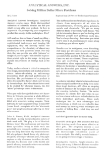
Can you guess what this microscopic picture is? Here are a few hints; it’s native to North America, plays an important role in many natural processes, and can be considered a sign of good luck or good fortune.
Still no clue? Here is a picture of the same subject on a regular scale.
If you guessed that it was some sort of plant, then congrats! But do you know exactly what kind of plant this is?
The plant in this picture is part of the genus Solidago, more commonly known as goldenrod. In other words, this is a microscopic picture of a weed. You might be wondering why we took the time to run microscopic scans of what is essentially the pesky plant version of a tumor that everyone takes extensive measures to keep out of their lawns. But goldenrod is actually highly misunderstood and carries a bad, not to mention false, reputation.
Although goldenrod is considered a weed in many regions in North America, it is actually just a harmless wildflower that is also the state flower of Kentucky and Nebraska. In Europe, goldenrod is considered a precious garden flower, and has also become common to the wild. In other regions, it is considered a sign of good luck. Many people believe that goldenrod causes hay fever in humans, but this is entirely false. Goldenrod inaccurately receives the blame for hay fever, when it is really ragweed that causes this, which just so happens to bloom around the same time as goldenrod. Although excessive handling of goldenrod can sometimes cause an allergic reaction, goldenrod is not linked to any typical allergies nor to hay fever. Actually, the pollen from goldenrod is too heavy to be blown by the wind, and it has to be pollinated mainly by insects. Honey from goldenrods is often dark and strong, and can sometimes have a spicy taste. Young goldenrod leaves are edible, and can also be used for herbal teas; the Native Americans used the seeds of goldenrod as food.
Still not convinced of the harmless, beneficial nature of this wildflower? Well how about the industrial uses of goldenrod, which includes being used for rubber in the tires of Model T Fords? Thomas Edison created a cultivation process that maximized the rubber content in goldenrod in order to produce plants that were 12 feet tall and up to 12% rubber. Henry Ford, who was dedicated to finding regenerative properties and alternative crops for materials for his cars, used goldenrod rubber for the tires in the Model T Ford that he gave to Edison as a gift. Later on, during World War II, extensive research and development was conducted to commercialize goldenrod as a source of rubber.
In addition to industrial uses, goldenrod has numerous medicinal uses. Historically, goldenrod was typically applied to the skin to help heal wounds and prevent infections, and Native Americans chewed on the leaves to relieve sore throats or toothaches. Goldenrod has been used to treat tuberculosis, diabetes, asthma, arthritis, and much more. Animal studies have shown that goldenrod can help reduce inflammation, relieve muscle spasms, fight infections and even lower blood pressure. With all of these beneficial side affects, it’s hard to deny the magical, medicinal powers of goldenrod.
Although many Americans consider goldenrod to be an annoying weed, it is actually a lovely wildflower with many practical, medicinal uses. Not only can goldenrod be used for rubber in car tires, but it can also help prevent infections and reduce inflammations. Not to mention, goldenrod does not cause allergies or hay fever. So, did all this information help to break the stigma about goldenrod? Hopefully it did!
Rockwell and Knoop microhardness testing are two methods of testing and documenting the hardness of materials, primarily metals. Rockwell is the “standard” test method and requires a ‘bulk’ sample or very thick layers (if tested in cross section). For thinner material layers and somewhat softer materials, Knoop testing uses a finer-point probe.
Durometer
is a method of testing the hardness of polymer materials. Manufacturing specifications for many structural plastics and other polymers require a specific Durometer (D).
Precision lapping and polishing
Mechanical polishing of samples in cross section is often needed to inspect or measure structures in semiconductor devices, or layers in plated or multi-layer materials. Samples are typically mounted in an epoxy and polished through a graded series of abrasive materials until a mirror-smooth surface is obtained. With our technology, we are able to achieve a precision lapped cross section through two (2) 25µm structures spaced approximately 25 mm apart.
Focused Ion Beam
Focused Ion Beam milling uses a gallium ion beam to mill into small (typically tens of microns across or less) sample regions of various materials to expose cross sections of the sample for imaging and critical dimension measurement, either in the FIB itself, using secondary electron and/or secondary ion imaging mode(s), or in the Ultra Field Emission SEM.
Broad Ion Beam
Broad Ion Beam milling employs three (3) argon (Ar) ion beams to mill cross sections through structures that are up to approximately 1.5 mm wide and up to approximately 0.5 mm deep, with FIB-quality results.
Cryomilling
At temperatures between -196° and -210° Celsius, liquid nitrogen makes many polymer and other soft materials brittle. Cryomillingis a preparation method used to mechanically break apart “bulk” polymer materials into finer grain materials or powders at liquid nitrogen temperatures to facilitate our analysis or subsequent processing by the client.
Ultramicrotomy/Microtomy
is a method for mechanically cutting thin or ultrathin sections of materials. This instrument is capable of slicing sections as thin as 40-60 nm for scanning transmission or transmission electron microscopy (STEM, TEM), or as thick as several microns for cross-sectional analysis by FTIR.


 Still not convinced of the harmless, beneficial nature of this wildflower? Well how about the industrial uses of goldenrod, which includes being used for rubber in the tires of Model T Fords? Thomas Edison created a cultivation process that maximized the rubber content in goldenrod in order to produce plants that were 12 feet tall and up to 12% rubber. Henry Ford, who was dedicated to finding regenerative properties and alternative crops for materials for his cars, used goldenrod rubber for the tires in the Model T Ford that he gave to Edison as a gift. Later on, during World War II, extensive research and development was conducted to commercialize goldenrod as a source of rubber.
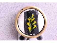 Animal studies have shown that goldenrod can help reduce inflammation, relieve muscle spasms, fight infections and even lower blood pressure. With all of these beneficial side affects, it’s hard to deny the magical, medicinal powers of goldenrod.